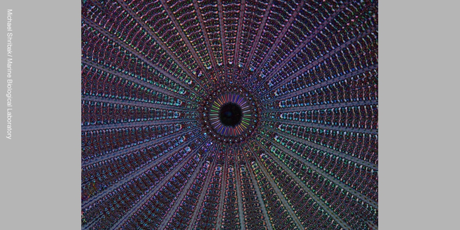
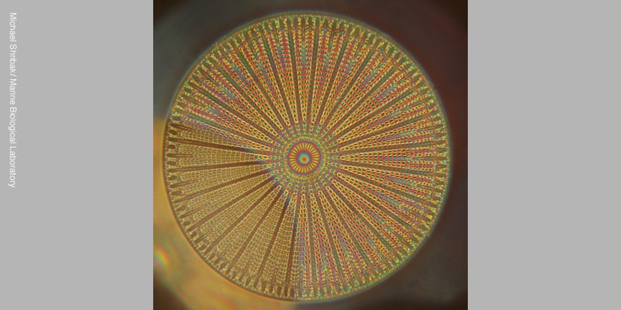
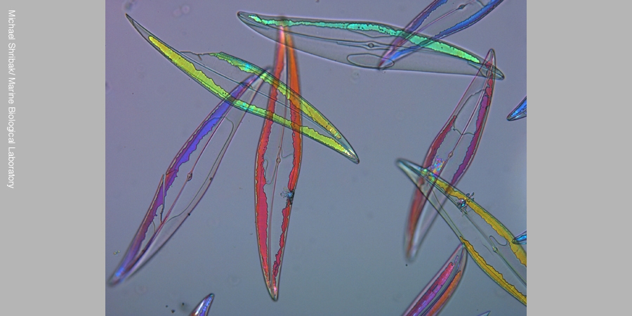
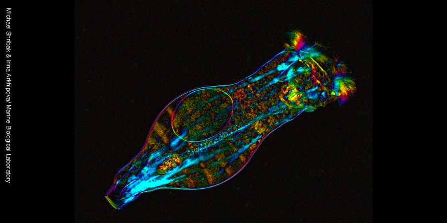
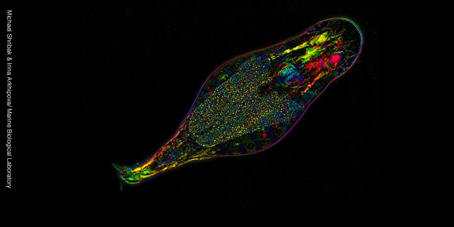
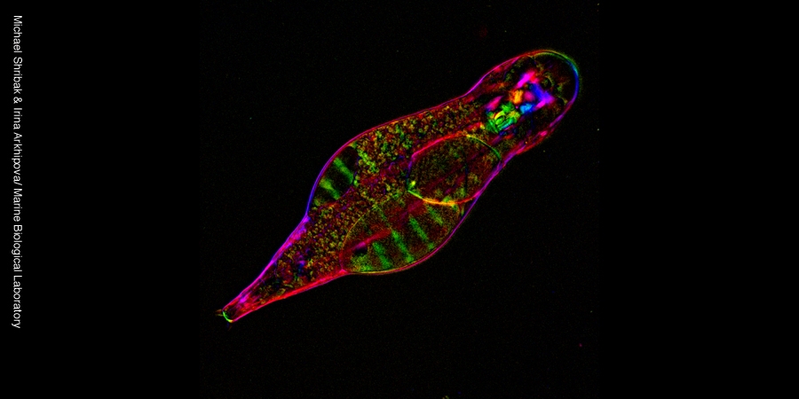
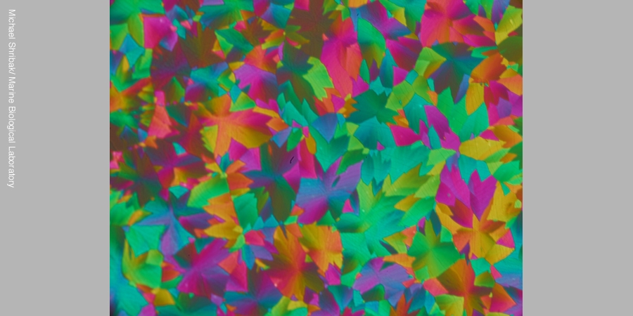
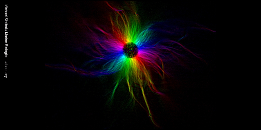
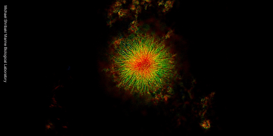
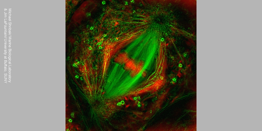
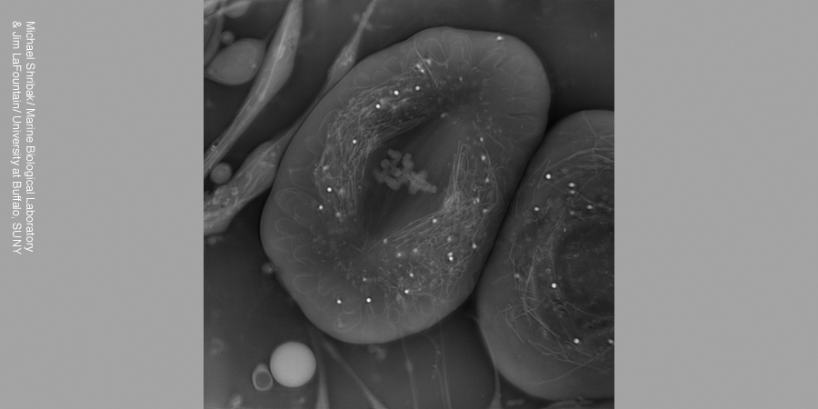
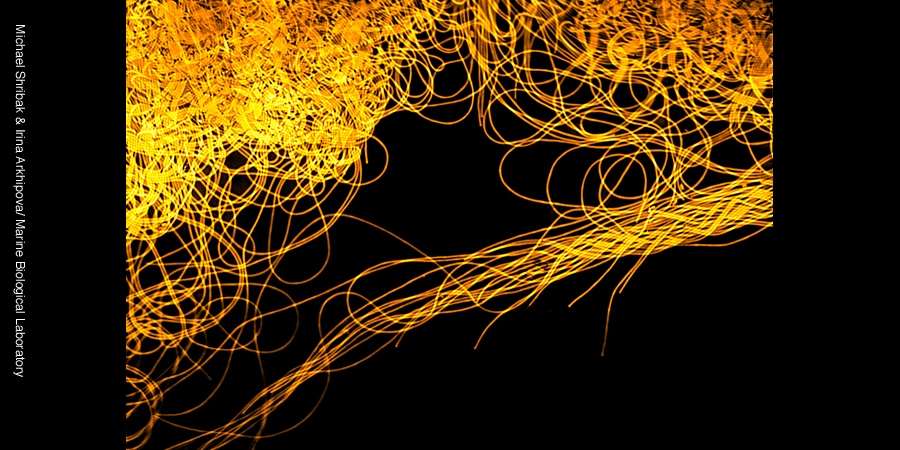
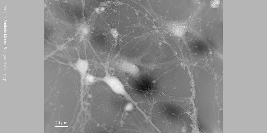
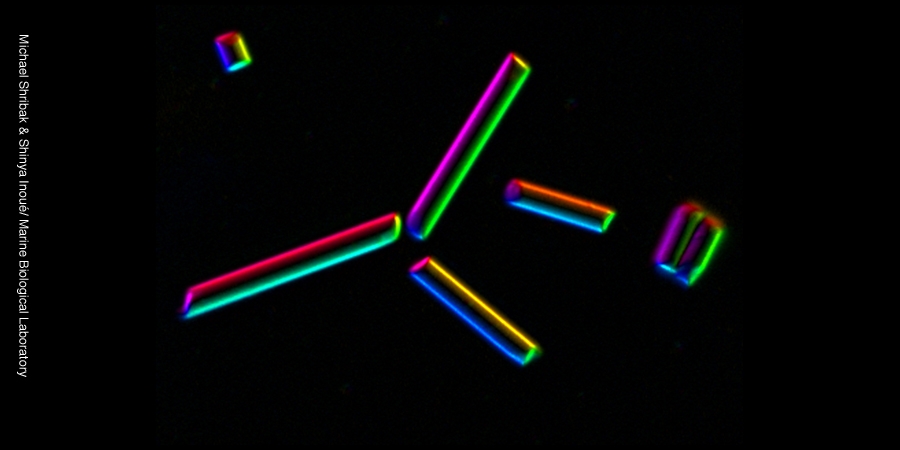
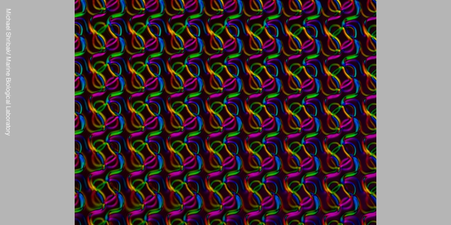
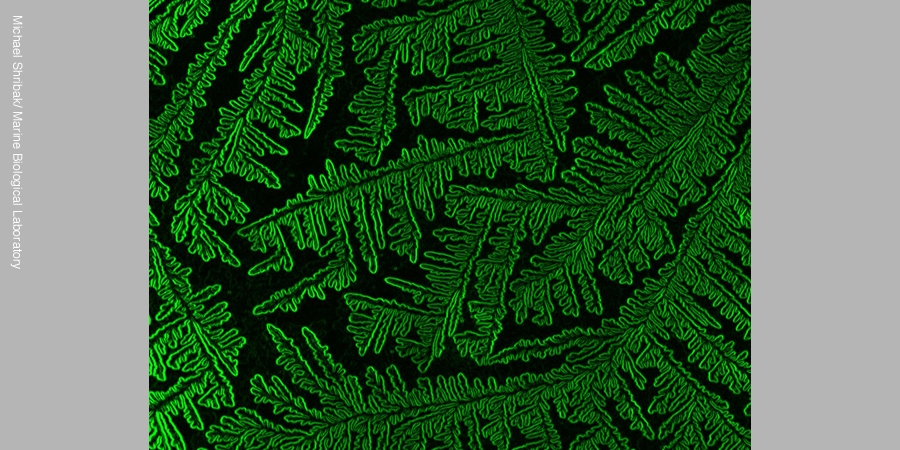
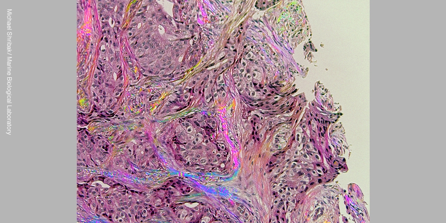
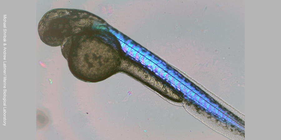
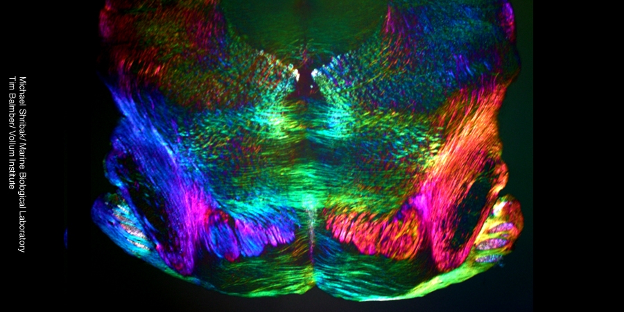
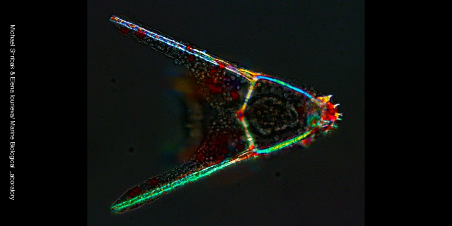
Back to Shribak Lab Shribak Lab Shribak Lab - Overview Shribak Gallery Home Groundbreaking Research At The MBL Research Centers Eugene Bell Center Faculty and Staff Shribak Lab Shribak Gallery Diatom Arachnoidiscus, birefringence image (polarized light microscope) Diatom Arachnoidiscus, interference image (Jamin-Lebedev microscope) Diatoms Pleurosigma, birefringence image (polarized light microscope) Rotifer Philodina roseola, birefringence image (polarized light microscope) Rotifer Adineta vaga, birefringence image (polarized light microscope) Rotifer Adineta vaga, birefringence image (polarized light microscope) Calcite crystal film, birefringence image (polarized light microscope) Aster consisting of microtubules (polarized light microscope) Aster, 3D orientation birefringence image (polarized light microscope) Crane Fly Nephrotoma suturalis spermatocyte, combined birefringence & phase images Crane Fly Nephrotoma suturalis spermatocyte, quantitative phase image (OI-DIC microscope) Bacterium Herpetosiphon aurantiacus (Chloroflexi), quantitative phase image (OI-DIC microscope) Neonatal Rat hippocampal neurons, ), quantitative phase image (OI-DIC microscope) 4µ-thick glass rods, quantitative phase gradient image (OI-DIC microscope) MFM diffraction grating, image size 90x67µm, quantitative phase gradient image (OI-DIC microscope) Drying water film, quantitative phase gradient image (OI-DIC microscope) Breast cancer tissue with collagenous stroma, birefringence image (polarized light microscope) Zebrafish embryo, birefringence image (polarized light microscope) Mouse coronal brain stem, birefringence image (polarized light microscope)