Volume Microscopy: from Array Tomography to PFIB SEM
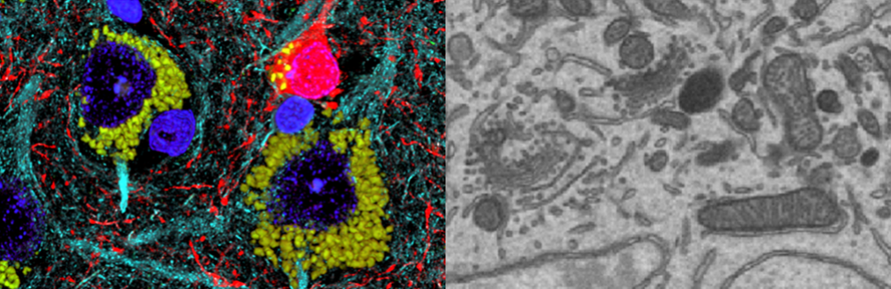
An intensive 6-day course on volume microscopy focusing on techniques ranging from array tomography (light or electron microscopy) to plasma FIB-SEM. This course will introduce participants into a dynamic area of technology development, at the forefront of 3D nanoscale and ultrastructural imaging that spans sub-organelle to cellular and tissue architectural scales.
Directors: Jemima Burden, University College London; Louis Kerr, MBL; Kristina Micheva, Stanford University; and Mark Terasaki, University of Connecticut
Course Description:
An intensive 6-day course on volume microscopy focusing on techniques ranging from array tomography (light or electron microscopy) to plasma FIB-SEM. This course will introduce participants into a dynamic area of technology development, at the forefront of 3D nanoscale and ultrastructural imaging that spans sub-organelle to cellular and tissue architectural scales.
Workflows range from relatively low-tech approaches, including array tomography for light and electron microscopy, to instrumentation-intense applications, such as plasma focused ion beam scanning electron microscopy (PFIB-SEM). The course program will focus on hands-on laboratory sessions and demonstrations, supplemented by background lectures delivered by leading scientists in this field. The faculty will represent a broad range of expertise to ensure that participants can experience many different volume microscopy approaches and test a broad range of instrumentation.
The course is aimed at graduate students and post-doctoral researchers but also core facility staff working in the biological sciences. It is limited to 12 participants to ensure ample hands-on time, personalized teaching and networking opportunities to explore the newest ideas surrounding the subcellular volume analysis of cells and tissues.
The course covers topics on:
- sample preparation for immunofluorescence array tomography, scanning electron microscopy (SEM) array tomography and PFIB-SEM
- serial ultramicrotomy, including traditional collection methods using rigid support and automated tape collecting ultramicrotomy (ATUM)
- fluorescence microscopy (epifluorescence, STED) and electron microscopy (SEM, PFIB-SEM, TEM) imaging of serial sections
- correlative light and electron microscopy by array tomography, pFIB SEM and ssTEM.
- image analysis for volume microscopy
The course emphasizes immersion and interactivity with lectures in the morning and complementary lab work in the afternoon/evening. With faculty provided samples, students will engage with real-life challenges and learn through problem-solving.