Three Embryology Course Students Win The Node's 2025 Image Contest

(Detail) This image of a mouse embryo, taken by MBL Embryology course students Nicole Roos and Anthony Wokasch, won the popular vote in the Node's 2025 microsocpy image contest.

The MBL’s 2025 Embryology course brought with it the much-anticipated return of a microscopy image contest sponsored by the Company of Biologists. The non-profit publisher and organization based in Cambridge, U.K., has interacted with the Embryology course since 1993 and previously sponsored contests of course images from 2011 to 2017. This year, the organization selected two images from 20 competitive submissions, which were showcased on its community site, The Node. The winning photos will be featured in the journal Development.

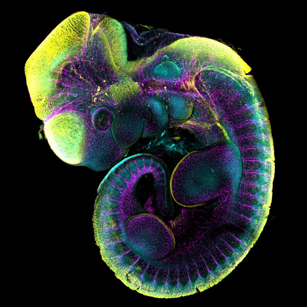
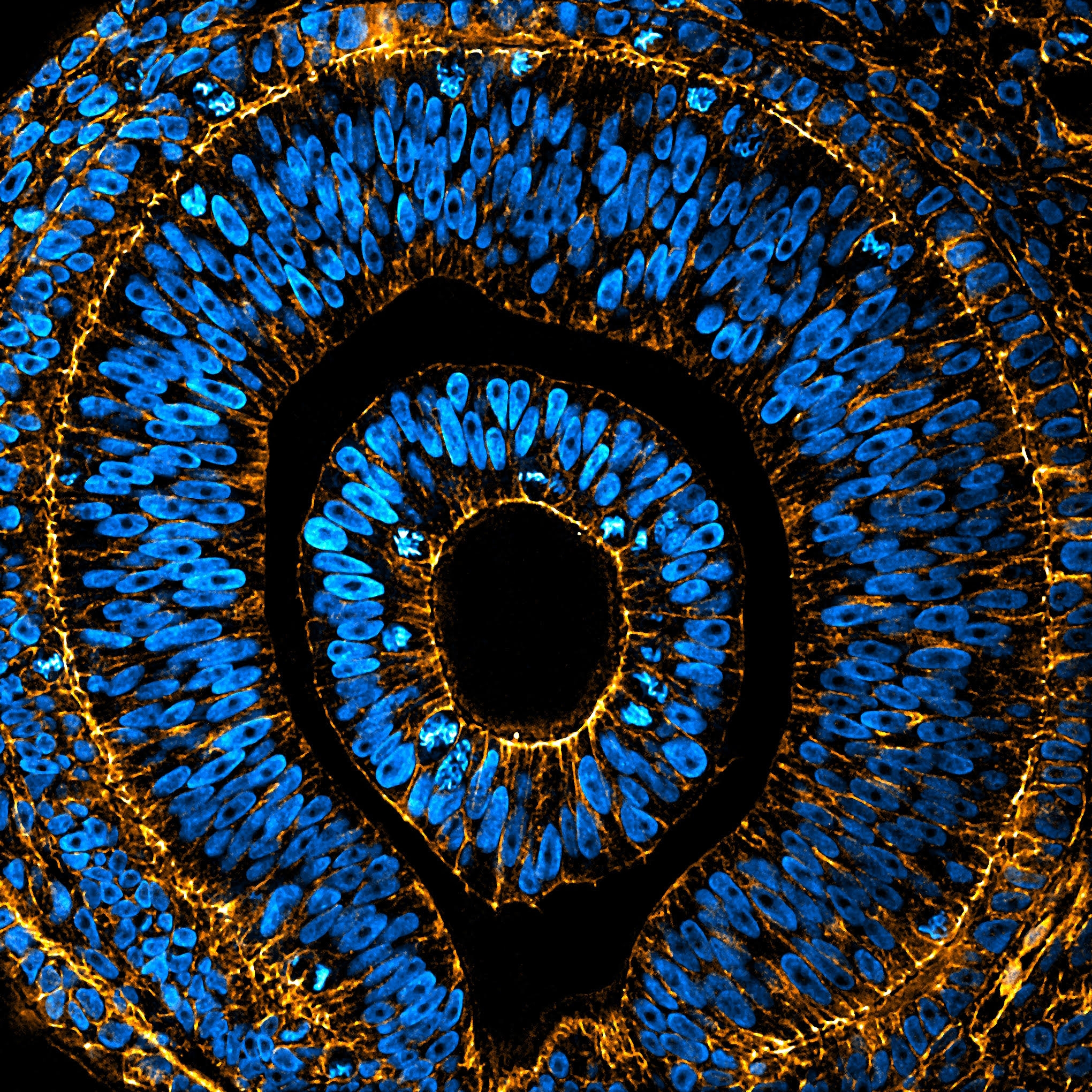
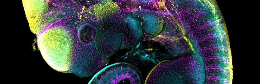
Embryology course students Nicole Roos (Emory University) and Anthony Wokasch (Vanderbilt School of Medicine) won the popular vote with an image of a mouse embryo. Arthur Boutillon (Technische Universität Dresden) had his editor’s choice winning photo of an anole lizard embryonic eye featured on the cover of the most recent issue of Development.

mouse embryo
The Node contest's winning image by popular vote. Mouse E10.5 embryo immunofluorescent staining of Sox9 (cyan), alpha-tubulin (yellow), and endomucin (magenta) protein. Image captured on Evident FV4000 point scanning confocal, lens UPLXAPO4X, na = 0.16, zoom = 1.04. Image processing conducted on Fiji. Credit: Nicole Roos and Anthony Wokasch / MBL Embryology course
lizard eye
The editor’s choice winning photo was featured on the cover of a recent issue of the journal Development. Embryonic eye of an Anole lizard stained for nuclei (DAPI, blue) and F-actin (Phalloidin, orange), imaged by spinning disc confocal microscopy and processed using ImageJ. Credit: Arthur Boutillon / MBL Embryology course
Remote video URL
This is one of two winning images in the The Node's 2025 photo contest of MBL Embryology course images. Image Credit: Nicole Roos and Anthony Wokasch/MBL Embryology course