MBL Scientists Discover Highly Organized Structures in Microbial Communities with Novel Imaging Approach

Contact: Diana Kenney, Marine Biological Laboratory
dkenney@mbl.edu; 508-289-7139
WOODS HOLE, Mass.-- Bacteria usually live in mixed communities with many different kinds of bacteria present. But it’s been largely unknown how these communities are organized, because the technology didn’t exist to see how they are structured in space.
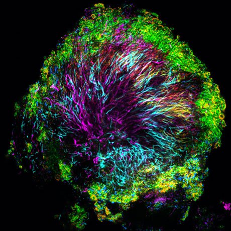
 A “hedgehog” structure in dental plaque, collected from a healthy volunteer using a toothpick. Corynebacteria, shown in magenta, form the core of the structure; other bacteria inhabit the structure at characteristic positions. Photo credit: Jessica Mark Welch, MBL
This week, for the first time, scientists describe distinct bacterial assemblages living in dental plaque, which they discovered using a novel imaging approach that “cuts through the overwhelming complexity of detail in microbial communities and allows common patterns to shine through.” The study appears in Proceedings of the National Academy of Sciences and was led by Jessica Mark Welch of the Marine Biological Laboratory (MBL), Woods Hole, and Gary Borisy of the Forsyth Institute, Cambridge.
Plaque on teeth, the team discovered, contains micron-scaled “hedgehog” structures in which eight different kinds of bacteria are radially arranged around a ninth kind, filamentous Corynebacteria. Seeing these structures offers scientists valuable information on how the bacterial members function that can’t be gleaned from genomic analysis, which specifies what microbes are present in a community, but not how they are organized.
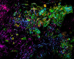
 A detail image of “corncobs” at the edge of a hedgehog structure. Each dot or rod is a bacterial cell. Photo credit: Jessica Mark Welch, MBL, and Blair Rossetti, Forsyth Institute.
“Microbes behave very differently depending on where they are and who they are next to,” Mark Welch says. “They will secrete entirely different sets of chemicals and metabolites depending on who their microbial neighbors are. So, if we want to accurately describe what these microbes are doing – really, what they are – we need to know where they are.”
The team proposes a model for how dental plaque develops, which is based on their imaging observations combined with plaque sequencing data from the Human Microbiome Project.
“This is a really exciting new way to look at microbial communities,” Mark Welch says of the spectral fluorescence imaging approach they developed at MBL. “The degree of organization we found in the hedgehog structure was amazing, as was the repeated finding of the same structure in different individuals. This finding that bacteria can develop such a degree of spatial organization may be generalizable to other microbiomes. We just have to go look.”
Citation:
Mark Welch JL, Rossetti BJ, Rieken CW, Dewhirst FE, and Borisy GG (2016) Biogeography of a Human Oral Microbiome at the Micron Scale. PNAS doi/10.1073/pnas.1522149113
####
The Marine Biological Laboratory (MBL) is dedicated to scientific discovery – exploring fundamental biology, understanding biodiversity and the environment, and informing the human condition through research and education. Founded in Woods Hole, Massachusetts in 1888, the MBL is a private, nonprofit institution and an affiliate of the University of Chicago.