MBL Scientist Takes a Closer Look at Human Microbiome | Falmouth Enterprise
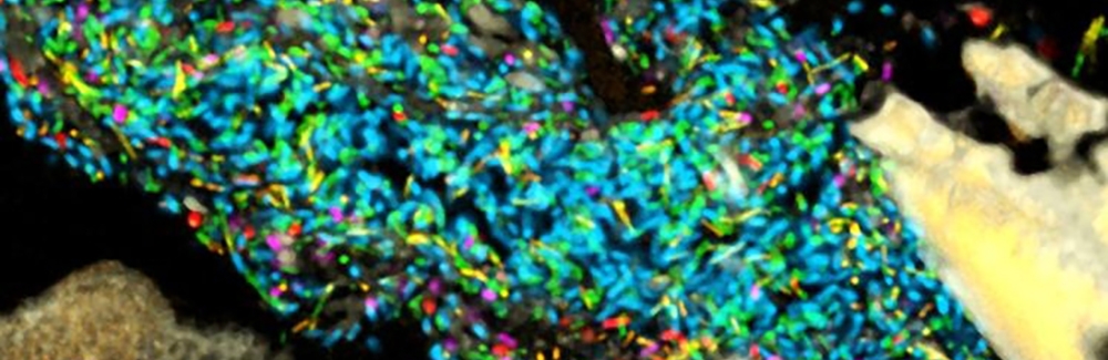
Long chains, coral branching, hedgehog and caterpillar shapes are some of the ways to describe the rainbow-hued images of microbial communities living in the body.
Scientists at the Marine Biological Laboratory are using fluorescent pictures to view these microbiome designs in the human mouth and gut. These patterns offer clues on why the microbes are there and what they may be doing.
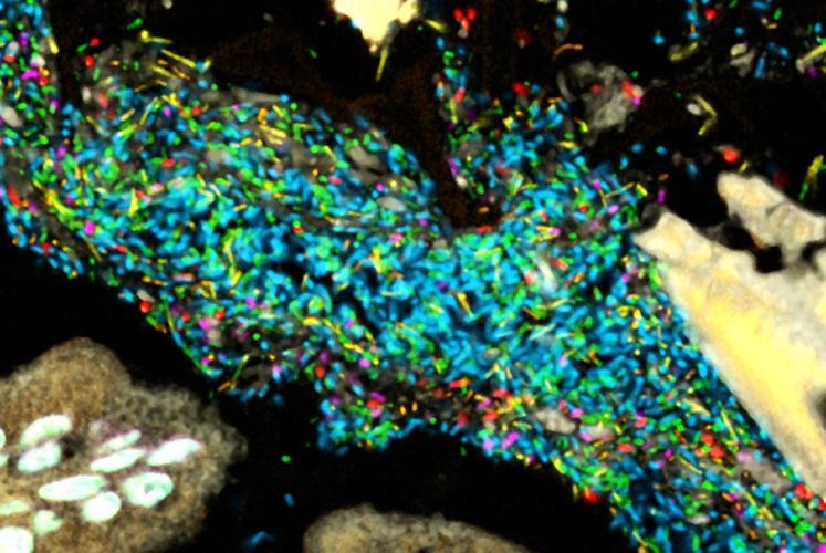
In a study published last month, scientist Jessica Mark Welch found that human gut bacteria appear happy to live side by side with other species rather than keeping to their own kind.
“It was interesting,” Dr. Mark Welch said. “We expected a lot more spatial organization and structure. What we saw was a lot of mixing. Most bacteria was next to a bacteria of a different type.” Read more of the article here.
Source: MBL Scientist Takes A Closer Look At The Human Microbiome | Falmouth News | capenews.net