A Cell Senses Its Curves: New Research from the Whitman Center

Can a cell sense its own shape? Working in the MBL’s Whitman Center research community, scientists from Dartmouth College developed an ingenious experiment to ask this question. Their conclusion – Yes – is detailed in a recent paper in the Journal of Cell Biology.
“Cells adopt diverse shapes that are related to how they function. We wondered if cells have the ability to perceive their own shapes, specifically, the curvature of the [cell] membrane,” says Drew Bridges, a Ph.D. candidate in the laboratory of Amy Gladfelter, associate professor of biological sciences at Dartmouth College and a scientist in the MBL’s Whitman Center.
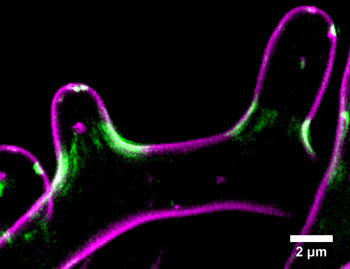
 The filamentous fungus Ashbya gossypii. The plasma membrane is visualized in magenta (FM-464), and the septin Cdc11a-GFP is in green. Credit: Andrew Bridges.
The team focused on the septins, proteins that are usually found near micron-scaled curves in the cell membrane, such as the furrow that marks where the cell will pinch together and divide. Using live-cell imaging at the MBL, they noticed that septins in a novel model system, the fungus Ashbya gossypii, tended to congregate on fungus branches where curvature was highest.
They then decided to recreate this natural phenomenon in the lab, using artificial materials they could measure more easily than living cells. Using precisely scaled glass beads coated with lipid membranes, they discovered that septin proteins preferred curves in the 1-3 micron range. They got the same result using human or fungal septins, suggesting that this phenomenon is evolutionarily conserved.
“This ability of septins to sense micron-scaled cell curvature provides cells with a previously unknown mechanism for organizing themselves,” Bridges says.
The idea for the glass bead experiment came from “many rich intellectual discussions with other members of the MBL community,” says Bridges, who has accompanied Gladfelter to the MBL each summer since 2012. “Both our collaborations and the imaging resources at MBL were central to this work.”
Citation:
Bridges, A. A., Jentzsch, M.S., Oakes, P.W., Occhipinti, P., and Gladfelter, A.S. (2016) Micron-scale plasma membrane curvature is recognized by the septin cytoskeleton. J Cell Biol 213:23-32, doi:10.1083/jcb.201512029.