Sea Stars Offer a Window into How Organs Take Shape
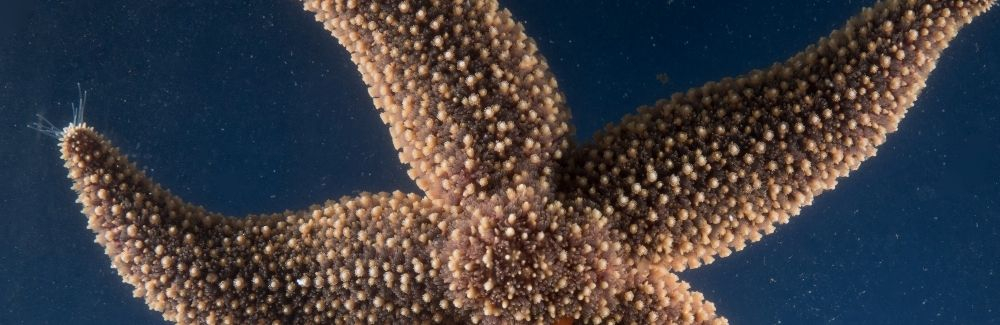
Biologists have long puzzled over how organs develop into their final shapes, and the nearly transparent bodies of young sea stars may offer a unique window into the organ development process.
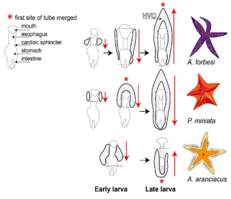
Many species of sea star develop a hollow tube during the larval stage that expands like a balloon in the body cavity, acting as precursor to their later organs. Margherita Perillo and her team at the Marine Biological Laboratory have named this tubal loop the hydro-vascular organ (HVO). Although the tube-shaped HVO appears deceptively simple, it shares a developmental feature with humans: our heart, lungs and kidneys also begin as simple tubes during embryonic development.
The larval sea star HVO begins as a small tube which will elongate, branch, and expand before acquiring its final shape. Perillo and her team are investigating the developmental processes that allow this transformation in the hopes of better understand how the blueprint for organ development is establised across species. Their previous findings, coupled with the fact that sea stars are among the closest invertebrate relatives to humans, make them an ideal candidate for the study of organ development. Sea stars “form embryonic organs like vertebrates do,” Perillo says, and their eggs and larvae are transparent, and abundant, allowing us to visualize the whole process.
By comparing HVO development across different species of sea stars like the Forbes sea star, bat sea star and red comb sea star, Perillo is answering the question of how the same organ develops across a group of animals.
While each of the species of sea stars studied has an HVO, they all develop a little differently. In Forbes sea stars, tubes first grow towards the stomach and then merge over the mouth, while in bat sea stars the first tubal connection is made early on before the tubes grow towards the stomach. In red comb stars the tubes merge near the intestine first. Despite these different developmental strategies, all the sea stars eventually end up with similarly shaped HVO’s.
So, what’s next? Now that her lab has laid the groundwork as to how HVO’s develop across sea star species, Perillo says that the next questions they hope to answer are “how we form organs starting from tubes, how these tubes maintain their proper orientation and shape, and once you have a fully grown, functional organ, how to prevent fibrosis in it.” By revealing how a simple tube develops into a functioning organ, the sea star HVO offers a powerful model for investigating these fundamental biological processes.