Our Ovary Blueprint is Ancient, According to Sea Stars
At first glance, bat sea stars, the nubbly, orange, many-footed creatures often found on the seafloor, seem about as far from humans as one can get. Appearances can be deceiving, however. Scientists this week published evidence showing human and sea star ovaries share similar genetics, cell types and signaling processes, despite their ancient evolutionary split.
Even so, the reproductive strategies of humans and sea stars could hardly be more different. Humans produce their lifetime supply of eggs while still embryos themselves. These egg cells remain dormant for years before maturing and being released from the ovaries after reproductive maturity is reached. Fertilization occurs internally, and this finite reserve of eggs gradually diminishes over time contributing to reproductive aging. In contrast, sea stars and their relatives can live for up to 200 years and continue producing millions of new eggs throughout their lives, releasing them into the sea for external fertilization.
Zak Swartz, an assistant scientist in the Eugene Bell Center at the Marine Biological Laboratory, has shown that despite these different reproductive strategies, bat sea star ovaries and human ovaries have cell types that express similar genes and reproductive signaling pathways in common. His group’s findings establish sea stars as a relevant model organism for studying stem cells and fertility treatments that could benefit human health. Their research also provides evidence that on an evolutionary level, some ovarian genes, cell types and mechanisms driving egg release were established in the last common ancestor shared by what are now bat sea stars and humans over 500 million years ago.
When Swartz began studying sea star reproduction, little was known about how the sea star ovary functions. His team set out to better understand “how the ovary contributes to reproductive health, and also to overall health as an endocrine organ that influences the whole body," he explained.
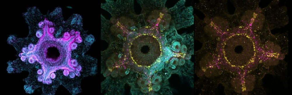
The scientists first needed to identify the cell types that make up the sea star ovary and understand how the ovary is organized. “The majority of animals reproduce through the fertilized egg, the only cell capable of making an entirely new body,” Swartz says. “That's pretty remarkable.”
In humans, each egg cell is surrounded by a layer of support cells called granulosa cells, which support the egg and keep it dormant until it receives signals to activate. Once these signals are received, the egg develops within a follicle and is eventually released for potential fertilization. Whether the support cells around sea star egg cells were similar to human’s granulosa cells, however, remained unknown.
Swartz and his colleagues were able to establish that bat sea star egg cells are surrounded by granulosa-like support cells. “From an evolutionary perspective, that suggests this cell type plays a fundamental role in reproduction,” Swartz says.
The team also found something unexpected when they took a closer look at the cells in the ovary: a network of what looked like interconnected neurons in the outer layers. This finding prompted the researchers to investigate which genes those cells were expressing and whether they might play a role in regulating reproduction.
Some of the genes looked familiar. The team identified signaling molecules that resemble neuropeptides involved in communication between the brain and reproductive organs in vertebrates, raising the possibility that the ovary itself has a built-in neuroendocrine system that helps regulate egg development and ovulation.
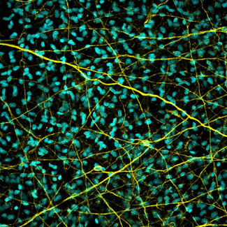
“There are lots of animals out there that don't have a brain but do have an ovary,” Swartz explained, so “how do they regulate reproduction in that kind of situation?” His team found evidence that the neurons inside the ovary may play a role in reproductive signals. Swartz suggests it’s possible that early in animal evolution, egg release depended on such a system before organisms became more complex and developed specialized structures such as brains.
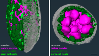
The cell organization of the sea star ovary may help explain how these animals continue producing eggs throughout their exceptionally long lives. “The ovaries are this big, branch-like structure, like a fractal, and really beautiful,” Swartz says. Inside these branches are clusters of cells that his team believes function as germline stem cells, continually replenishing the ovary's supply of oocytes.
His team hypothesizes that these cells follow a different developmental path than their counterparts in mammals. In mammals, a founder cell produces an egg and is depleted in the process. In sea stars, however, the same type of cell may be capable of both producing eggs and renewing itself, allowing the ovary to maintain egg production throughout the sea star's life.
Now that Swartz and his team have identified the major cell types in the ovary, he hopes to understand how those cells communicate, particularly, how the stem cell population is maintained over time.
“Since sea stars, genetically, have a pretty similar toolkit of genes that we have as humans,” they must be turning on or off “those same genes in a very different way that enables long-term stem cells to persist.”
Understanding these genetic shifts may be therapeutically useful for humans. Swartz envisions a world where a better understanding of bat sea star stem cells may enable us to develop therapies for humans that aid in restoring fertility or could be used to augment stem cell therapies.