MBL Scientists Describe Major Differences Between Related Desert and Aquatic Algae
By Stephanie M. McPherson
Staying alive in the desert is no simple matter for green algae whose evolutionary ancestors lived in the ocean. How can some algal species survive extreme drought, while others desiccate and die? Understanding this difference can provide important information on requirements for drought tolerance that it may be possible to apply to larger plants as the climate changes.
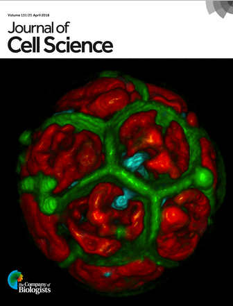 Journal cover photo (above): Sixteen daughter cells still packed within the mother cell wall of a desiccation-tolerant green microalga Acutodesmus bajacalifornicus. Volume was rendered from confocal z-stacks and shows daughter cell membranes (green), nuclei (blue) and chloroplasts (red) after multiple fission of the single mother cell. Z. Cardon et al.
Researchers have described a new genetic model to study exactly this question in a paper published in Journal of Cell Science. In a group of five closely related species of green algae called Scenedesmaceae, three that have adapted to life in desert crust can withstand multiple rounds of desiccation, whereas their two aquatic cousins perish after drying out once. The paper also details major reproductive differences between the two groups.
“How can that be?” asks MBL Senior Scientist Zoe Cardon. “What in the genetic makeup of these species is making such a difference for both their form and their function?”
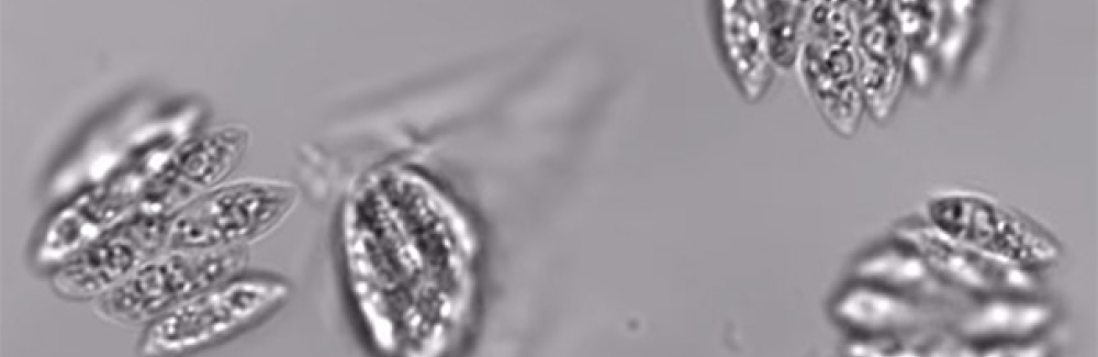
Cardon brought these microbes into her lab at the MBL, and she and Research Scientist Elena Lopez Peredo either dried them out rapidly, using airflow through a chemical hood, or more gradually at the bench. They then observed the effect of desiccation on each species. They saw that after the aquatic microbes had dried, no amount of water could revive them. The desert microbes, however, showed evidence of active photosynthesis after multiple rounds of desiccation and rehydration.
“As they dry out, you can see the photosynthesis just shutting down,” says Cardon. “You add a little more water and then, poof! It’s off to the races again.”
The researchers also noticed a striking difference in the way these two genetically-similar organisms reproduce. Joining forces with MBL Whitman Center Scientist Magdalena Bezanilla of Dartmouth College, the paper also compares for the first time the microbes’ reproductive processes. The aquatic microbes fit up to 16 daughter cells within the walls of one mother cell, while the desert microbes can pack in up to 64.
“It almost looks like all those facets on a soccer ball,” says Cardon. “Except imagine that all those seams on the soccer ball go into the center of the sphere.”
The desert algae cells spill their daughter cells out to live individual lives. But the aquatic algae release daughter cells en masse, joined in a flat sheet.
“Those daughter cells are stuck together permanently,” she says. “They pop out like a little colony, all at once in a big sheet.”
Follow-up studies will investigate the activation of genes that cause the major differences in the reproductive behavior of such closely related species.
This family of algae was not completely unknown before this work. The aquatic species are used in biofuels development, and Cardon hopes further exploration of these microbes’ functions will help improve those efforts. In the long-term, understanding the genetic differences between these organisms could provide more information on how to bolster the drought resistance of agriculturally important plants and other larger-scale greenery.
Cardon, an expert on photosynthesis, credits the MBL for making possible the collaboration with Benzanilla, who focuses on microscopy and cell division.
“This would be impossible by myself,” says Cardon. “[Benzanilla] comes every summer and we’re able to do this research together with the incredible microscopes and facilities of the MBL.”
Citation:
Cardon, ZG et al (2018) A model suite of green algae within the Scenedesmaceae for investigating contrasting desiccation tolerance and morphology. J. Cell Science, doi: 10.1242/jcs.212233.
J. Cell Science Research Highlight: http://jcs.biologists.org/content/131/7/e0703
—###—
The Marine Biological Laboratory (MBL) is dedicated to scientific discovery – exploring fundamental biology, understanding marine biodiversity and the environment, and informing the human condition through research and education. Founded in Woods Hole, Massachusetts in 1888, the MBL is a private, nonprofit institution and an affiliate of the University of Chicago.