The MBL in Living Color: Selected Microscopy Images and Movies

If you spend any time at the MBL, you'll soon be dazzled by the images and movies that stream from the labs and courses. Scientists here continually push the boundaries of speed, resolution, and general awesomeness in helping us visualize life at the molecular, cellular, and organismal levels of action. Here is a small sample of images and movies generated by MBL scientists, faculty and students that bubbled into our news feed in 2018. Here's to a bright New Year!
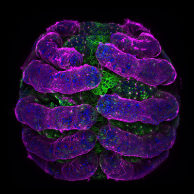 Spider embryo imaged by Tessa Montague in the MBL Embryology Course. This image took 5th place in Nikon's 2018 Small World Photography Competition.
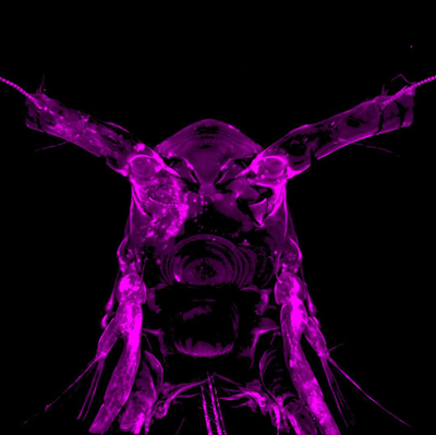 Copepod encased in a hydrogel. Credit: MBL Research Scientist Eric Edsinger
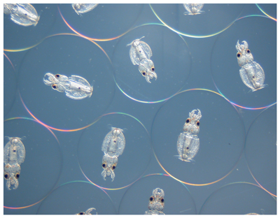 Squid embryos fertilized and cultured in vitro. Credit: MBL Whitman Fellow Karen Crawford, St. Mary’s College of Maryland
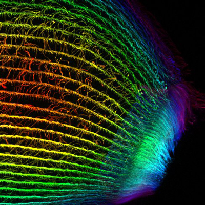 Cilia on the single-celled organism Stentor. Credit: Aidan Fenix, MBL Physiology Course
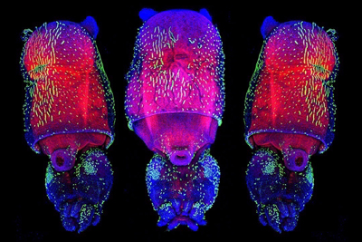 Juvenile squid. Credit: Wang Chi Lau, MBL Embryology Course. Science magazine selected this as one of its favorite published images in 2018.
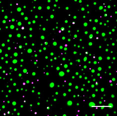 Distinct protein droplets form with different RNA. Credit: E.M. Langdon et al., Science, 2018
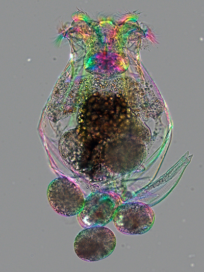 Female rotifer. Credit: MBL scientists Michael Shribak and Kristin Gribble
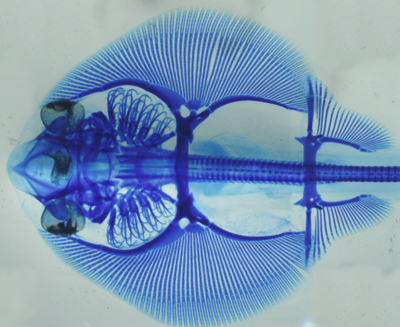 Cartilage staining of little skate embryo. Credit: MBL Whitman Fellows J.L. Gomez-Skarmeta and Tetsuya Nakamura
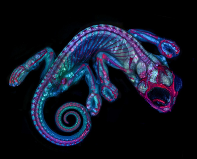 Chameleon embryo imaged by Teresa Zogoda in the MBL Embryology Course. This image received an honorable mention in the 2018 Nikon Small World Competition.