The Inner Ear in Living Color | @m_scheibinger
Postdoctoral scientist Mirko Scheibinger of Stanford University School of Medicine, a teaching assistant for the MBL Biology of the Inner Ear (BIE) course, has been Tweeting microscopy images from the course, each one more beautiful than the next.
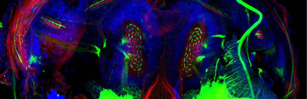
 Mouse inner ear spiral ganglia culture. Image taken with Zeiss LSM710 in Biology of Inner Ear course 2017. Courtesy of Mirko Scheibinger
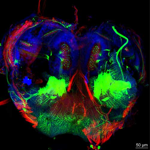
The course focuses on the hard-to-access but vital sense organs in the inner ear, which are essential for detecting sound as well as head and body movements that provide equilibrium and spatial orientation to animals as they navigate their environment. Currently co-directed by Paul Fuchs, Johns Hopkins University School of Medicine, and Stefan Heller, Stanford University School of Medicine, BIE takes a comparative approach and studies the inner ear systems of many animals, from fish to mammals. As the images below vividly illustrate, the course has an emphasis on high-resolution fluorescence microscopy of inner ear tissues.
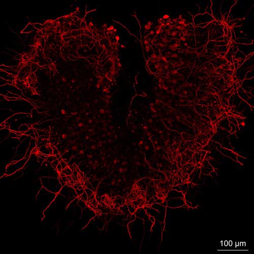 "Neurons in Love" - In vitro culture of spiral ganglion neurons from the cochlea of the inner ear. Image taken with Zeiss LSM880 with Airyscan in BIE course 2017. Courtesy of Mirko Scheibinger
"We cultured these neurons for one day and labeled them with an antibody against Tuj1, which is known to label neurons. These neurons randomly grew into a heart shape. Very cool!" Scheibinger says.
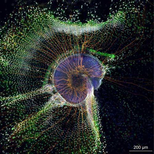
 Squid statocyst (inner ear sense organ in some aquatic invertebrates). Image taken with Zeiss LSM710 in BIE course 2017. Courtesy of Mirko Scheibinger