The Science Behind The Art Of Nikon’s Small World Competition | Forbes

Nikon recently announced the winners of their annual Small World photomicrography competition. It’s a contest open to any photos taken with a microscope. The finalists are usually intriguing and surprising images of the natural world, shown in a way that most people haven’t seen before.
But since taking such images is reserved for people with access to scientific microscopes, they’re often made by scientists, in the context of a research experiment or training course. Often the initial goal of these images is to make new discoveries, rather than create art. It just so happens that certain methods of studying nature create really nice images.
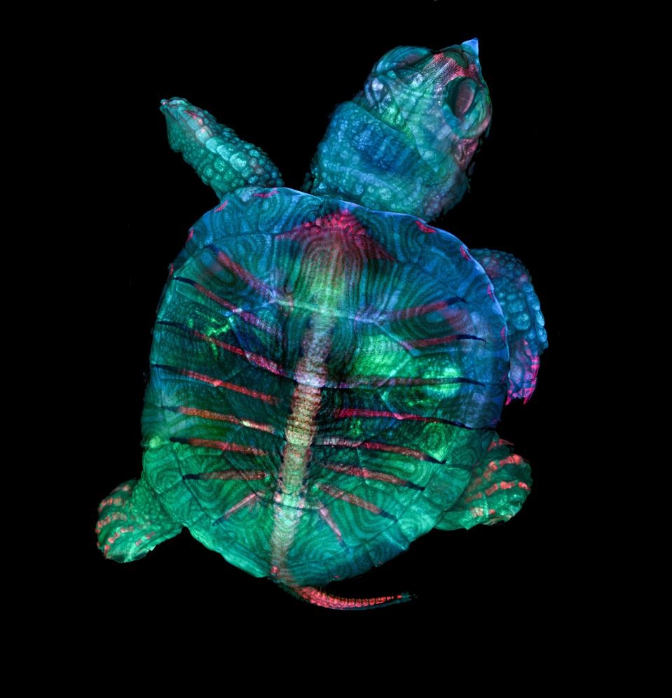
This year the first prize went to a fluorescence image of a turtle embryo, taken at the Marine Biological Laboratory in Woods Hole, Massachusetts by Teresa Zgoda and Teresa Kugler. They were both interns at Zeiss at the time, and took part in the 2017 MBL embryology course. This course is organised every year for biologists who study animal development in different species. Usually, when researchers learn microscopy techniques they visualize cells or tissues, so taking a picture of an entire organism (even a tiny one) requires some different skills. Read more of the article here.
Source: The Science Behind The Art Of Nikon’s Small World Competition