Light Microscopy Bootcamp Opens New Worlds for PhD Student Researchers
It can take some tinkering to find the ideal recipe for a new research training course at MBL, as Prof. Ed Munro of University of Chicago is finding with the Light Microscopy Bootcamp.
Munro directed the bootcamp for the second time last fall, and it became clear that students bringing in specimens from their home labs is “hugely successful.”
“Several students wanted to figure out how to do better microscopy to answer the specific questions they are working on [at home]. And the course opened up possibilities for them that they didn’t even know existed. Some students are acquiring images they didn’t even know they could acquire, and sending them back to their labs. And they are really, really excited about that,” Monro said. “It’s something we will do in an even more deliberate way in the future.”
“They can’t wait to go to the next lab meeting at their organization, to show their colleagues what microscopy techniques they could be using,” said Noah Mitchell, assistant professor at UChicago who led the course with Munro.
The two-week bootcamp was first held at MBL in 2023 for UChicago graduate students. This year, the program also accepted students from other institutions.
“We select students for whom this could really be a transformative experience,” Munro said – those who envision microscopy as a fundamental tool for their PhD research.
The course is structured like the MBL’s Advanced Research Training Courses, with lectures in the morning and lab work in the afternoon. The first week, students built a simple compound microscope and learned the fundamentals of optics on a variety of advanced microscopes at the MBL.
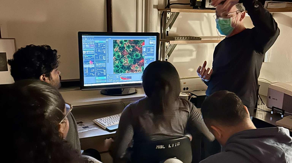
Bootcamp faculty Rick Fehon of UChicago (standing) imaging cell division in the larval fly brain on the Zeiss LSM 780 with students Pavan Rao, Trinity-Paris Foster, Amina Bradley, and Christian D. Del Valle Colón. Credit: Tiffany Lu
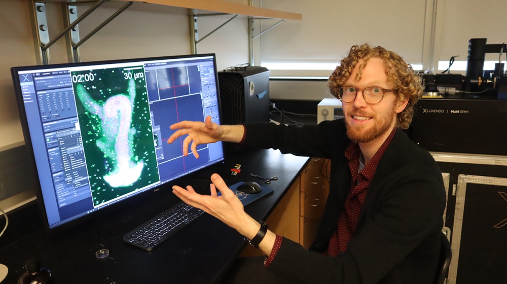
Bootcamp faculty Noah Mitchell of UChicago trained students in advanced light-sheet microscopy on a loaned Bruker Luxendo MuVi SPIM. Credit: Diana Kenney
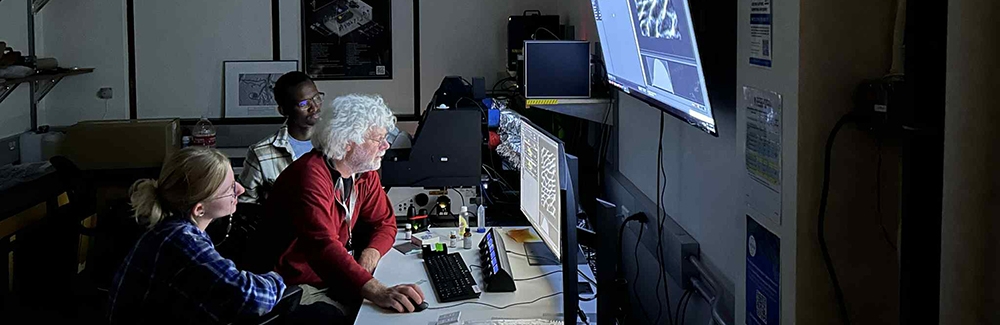
MBL Research Scientist Matthew Parent trains students Erick Torres-Guasp (left) and Amina Bradley (right) on a custom dual-view line-scanning confocal microscope in the MBL’s Imaging Innovation Center. Credit: Diana Kenney
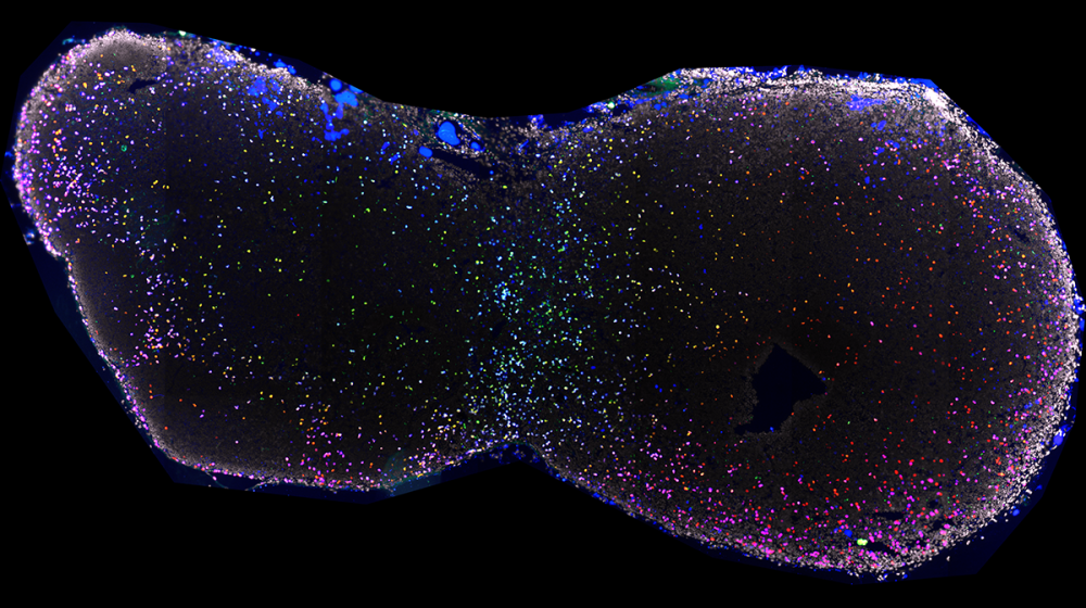
Mouse cervical lymph node stained for nuclei (DAPI, white), IL17+ cells (red), IFN+ (green), and T cells (CD3, blue). Image taken on Zeiss LSM 780 with supervision from teaching assistants Sarah Christensen, Isaak Tarampoulous, and Tiffany Lu. Credit: Emily McNaughton (2025 Light Microscopy Bootcamp student).
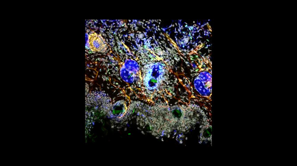
Mouse ear skin stained for nuclei (DAPI, white), Schwann cells (Plp1, red), CCR6+ B and T cells (green), and T cells (CD3, blue). Image taken on Leica Stellaris 8 with supervision from teaching assistants Sarah Christensen, Isaak Tarampoulous, and Tiffany Lu. Credit: Emily McNaughton (2025 Light Microscopy Bootcamp student)
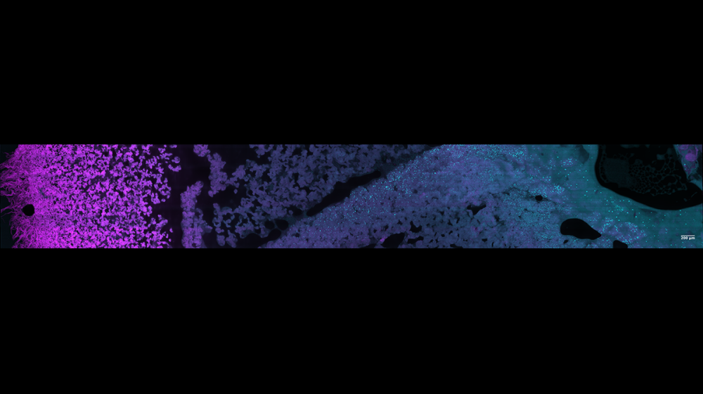
Bacillus subtilis colony biofilm with labeled Ypet (magenta) and antibiotic protein Subtilosin-A (cyan). Image taken on dual view line-scanning confocal with supervision from Matthew Parent. Credit: Amina Bradley (2025 Light Microscopy Bootcamp student)
“We are building up, day by day, the conceptual framework for thinking about how to achieve optimal resolution on different microscopes,” Monro said. “How do you [assess] the trade-offs between spatial, optical, and temporal resolution and signal-to-noise? The fundamental concepts of good microscopy run as a thread through the whole course.”
In the second week, students split into small groups and explore a particular area more deeply, such as single-molecule imaging or cutting-edge light sheet microscopy. “In practice, some students are bouncing from one group to another, because they don’t want to restrict themselves,” Munro said.
Or the students can image specimens they brought from home in different ways. “That has worked really well. We had to be more flexible, but we had teaching assistants who took the course before who facilitated that flexibility,” Munro says. “They did a fantastic job. I think it’s a good model to draw on course alumni to serve as TAs.”
“There are also students who are really excited to work with an organism they’ve never seen before,” Munro said. MBL scientist Zak Swartz, for example, spent two days imaging sea star embryos with the students. “They were up at midnight last night trying to figure out, how many ways can you image these embryos?” Munro said.
Munro and Mitchell look forward to the next iteration of the course, with ideas to continuously improve. One thing is for sure: Imaging a variety of organisms is a hit.
Other faculty who lectured or taught imaging techniques in the 2025 Light Microscopy Bootcamp included, from MBL, Rudolf Oldenbourg, Matthew Parent, Nipam Patel, Zak Swartz, Riley Walsh and Carsten Wolff, and from UChicago, Rick Fehon and Patrick LaRivière.