Central Command: Injury to Axolotl Tail Activates Distant Neurons in Brain, Promoting Regeneration
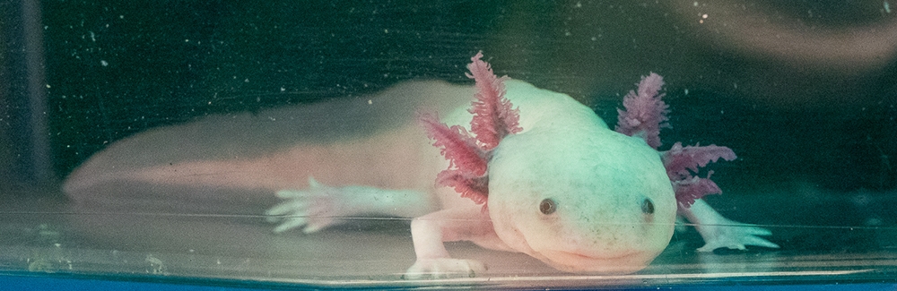
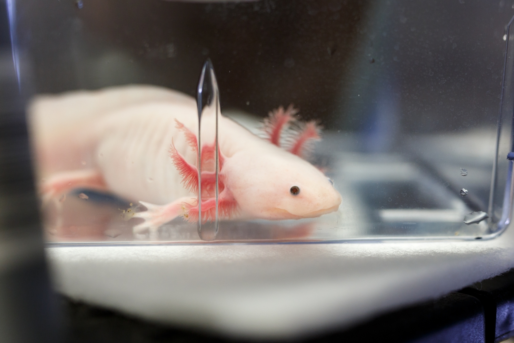
WOODS HOLE, Mass. — The axolotl is renowned for its extensive ability to regenerate organs and body parts, including its spinal cord. Studies on spinal cord regeneration, however, have focused on axolotl cells next to an injury site, leaving the brain’s role in regeneration a relative mystery.
A new study from researchers at the Marine Biological Laboratory (MBL), Woods Hole, reveals that activating a specific group of neurons in the axolotl brain is essential for tail regeneration.
Their findings point to the possibility that a comparable group of neurons impacts regenerative responses in mammals. The study, led by MBL Associate Scientist Karen Echeverri, was published last week in npj Regenerative Medicine.
“Sometimes we think about injury and regeneration as just the response locally at the injury site, what is happening in the cells there, and we forget that everything in our body is actually controlled by our brain,” Echeverri says. “What happens in our brain might be the difference between what happens in a human in tissue that regenerates, like the liver, or doesn't regenerate, like most other organs.”
Decoding the Brain After Injury
Previous research on the axolotl brain has characterized its cell types, but not which cells are activated in response to injury elsewhere in the body, Echeverri says. The new study dives deep into the activity of a specific group of neurons that extend axons from the telencephalon — an area near the front of the axolotl brain — into the hypothalamus, a region near the base.
When activated, a protein called extracellular-regulated kinase, or Erk, sets off molecular chain-reactions that cause changes in gene expression. Echeverri and collaborators have previously found Erk levels increased in nervous system support cells, called glia, in the spinal cord after injury.
This time, the researchers found Erk activity increased in the group of telencephalon neurons after several types of injuries, including tail and limb amputation. Blocking Erk from working normally in the brain resulted in significantly shorter regenerated tails.
In the hypothalamus, the neurons increased production of a protein called neurotensin, which the researchers also found to be important for regeneration; like Erk, when they blocked neurotensin from working, axolotl tails regrew to significantly shorter lengths.
The neurons first caught Echeverri’s attention when she was an assistant professor at the University of Minnesota, studying how Erk functions in the spinal cord after injury. One of her students, Keith Sabin, looked for Erk in the axolotl brain and found it in the telencephalon neurons.
Echeverri then picked the neuron thread back up when postdoctoral fellow Sarah Walker — the paper’s first author, now of Brock University — joined her lab at the MBL in 2020.
The pair now hope to investigate if the same group of neurons is activated in mammalian brains following injury. They’re also aiming to identify the key molecules that travel between the injury site and the brain and determine how the brain responds to different types of injuries.
“How does it understand that it’s injured its spinal cord versus it’s injured its limb? In our paper, we compared a limb injury versus a tail amputation, and we see that the same area of neurons is activated,” Echeverri says. “But we need to now delve much more into that data to see if there's a subpopulation of activated neurons that is specific to the limb.”
Humans can regenerate parts of their bodies, but only certain tissues like skin, muscles, and the liver. But even if our regenerative abilities were akin to an axolotl’s, it would take us significantly longer to regenerate after an injury to the spinal cord, Echeverri says, simply because we're much larger organisms.
During a long regeneration, humans could be more susceptible to infection and even injure ourselves further by trying to move around. It’s possible that we instead evolved to heal wounds and generate scar tissue to avoid these outcomes, Echeverri says.
“That's one theory, since we do have some capacity to regenerate,” she says. “Question is, can we harness that capacity and get it to regenerate much faster?”
Citation: SE Walker et al. (2025) Neuronal activation in the axolotl brain promotes tail regeneration. npj Regenerative Medicine, DOI: 10.1038/s41536-025-00413-2
Collaborating institution: Translational and Functional Genomics Branch, National Human Genome Research Institute, National Institutes of Health
—###—
The Marine Biological Laboratory (MBL) is dedicated to scientific discovery – exploring fundamental biology, understanding marine biodiversity and the environment, and informing the human condition through research and education. Founded in Woods Hole, Massachusetts in 1888, the MBL is a private, nonprofit institution and an affiliate of the University of Chicago.